


One of the mods mentioned that it might be a good idea to have a bunch of nasty pictures of rabbit diseases so I figured I could do that and it also might help some people to identify what is wrong with their rabbit(s) before something really bad happens. If this needs to be deleted please do so.
The information for these diseases came from the following websites. All credit goes to them. Visit their websites for more detailed information as this is just a VERY cursory look at some of the diseases.
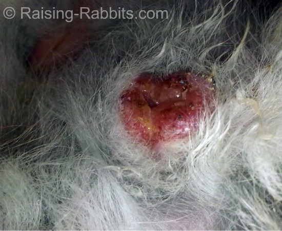
http://www.raising-rabbits.com/rabbit-diseases.html
http://www.lbah.com/word/rabbit-diseases/
http://www.vetnext.com/search.php?s=aan ... 77559%2080
http://www.cottontails-rescue.org.uk/gutstasis.asp,
http://www.cfsph.iastate.edu/DiseaseInf ... ic-disease,
http://www.freedomforfarmedrabbits.com/ ... sease.html,
Otherwise known as Snuffles
Pasteurella multocida is a nasty little germ. When a rabbit comes down with symptoms from this bacterium, the vet will call this disease Pasteurellosis, the general term used when the rabbit’s immune system has been overwhelmed with Pasteurella multocida.
The leading researchers believe that 100% of rabbits, or at least 100% of rabbitries, have been exposed to pasteurella (Rabbit Production, Sixth Edition, pg 213). The germ is everywhere in the rabbit world; the best protection is boosting your rabbit's overall health.
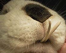
This bacterium is the cause of the respiratory condition known as "snuffles." Snuffles actually describes a symptom, not exactly the illness. Snuffles are paroxysms of head-jerking sneezing that occur on a regular basis. The symptom of snuffles is almost certainly a result of pasteurella multocida. If your rabbit is sneezing, and you see ANY white sticky snot in its nose, it’s not a cold, it’s Pasteurella multocida.
Picture all those little green mucus critters on TV moving into your rabbit's lungs, suitcases and all, but with NO REMEDY.
This picture of a snotty rabbit nose is published by Tufts University.
Listen to the rabbit experts: "There’s no such thing as a ‘cold’ in rabbits," is the unequivocal statement in Rabbit Production (p. 213). "Mucopurulent nasal discharges (pus) that many people attribute to colds are almost invariably caused by P. multocida in conjunction with another bacterium called Bordatella bronchisepticum. This is not a transient condition like a cold, but one with permanent adverse effects on the animal."
Before we assume rabbit-sneezing is due to pasteurella, check to ensure the rabbit is not sneezing from environmental causes:
feed dust sniffed by the rabbit - the sneeze will be short and dry.
Drinking water in the nose - the nose might be damp on the outside tip of the nose, and the sneeze will be brief.
In the case of pasteurella: The rabbit keeps on sneezing on and off, and you notice matting on the insides of the front legs from wiping the white snot away with the front legs (they haven't figured out how to use tissues), and especially, you actually see white snot bubbles as the rabbit sneezes. Thick white snot is pasteurella multocida.
A Whole Repertoire of Trouble with Pasteurella Multocida.
Snuffles easily turns into:
Pneumonia
Metritis - infection in doe’s womb
Orchitis - infection in buck’s testicles
Colds - there are no colds, only pasteurella multocida (99% of the time)
Wry Neck - middle or inner ear infection
Weepy Eyes - eye infection closing off the tear ducts
Body abscesses - marble-sized to golf-ball sized boils, filled with thick white cheesy pus
Body ulcers - ulcerations, on skin, tongue, eyes, other locations
Bone infections - frequently associated with infected teeth
Infections in roots of teeth
I doubt this list is complete...oh yeah, death...
Sometimes the nasal discharge is so chronic that the fur is actually missing.
Other respiratory signs of Pasteurella include sneezing, congestion, and conjunctivitis. The tear ducts (lacrimal ducts) can become clogged with dried discharge, causing excess tearing and subsequent scalding of the skin around the eyes and face.
In addition to the respiratory tract, the bacteria can also infect the reproductive tract, the sinuses, the eyes, the ears, and the internal organs. It sometimes causes abscesses under the skin. These abscesses can become chronic and require surgery to correct. Severe cases can cause central nervous system symptoms like oscillations of the eyes (nystagmus), circling to one side, and severe tilting (wry neck or torticollis) of the head.
This rabbit has a neurologic problem from Pasteurella
Rabbits with ear infections might paw at the ears and those with internal organ infections might have poor appetites and lose weight. If the reproductive tract is infected discharge is commonly noted.
The following sections contain graphic surgical pictures, and may not be appropriate for everyone.
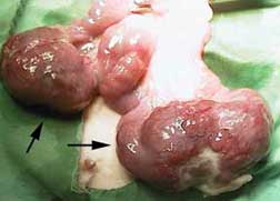
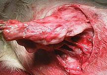
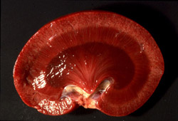
This is a healthy uterus during a routine spay (OVH). The healthy pink uterine horns are easily seen (white arrow).
The arrows point to the typical appearance of a uterus infected with Pasteurella. Cancer can also look like this.
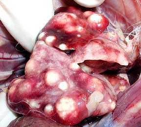
This is what these lungs could look like on an autopsy. All the white spots correspond to the white spots on the radiograph above.
Rabbits have a very thick and tenacious discharge when they form an abscess, and require more care than the abscesses of most other animals. Surgical removal can be difficult, especially in the chronic cases, because the abscessed area can become extensive in nature. Multiple surgeries might be needed, and wound care at home is necessary.
This is a severe abscess on the back of a rabbit that has been anesthetized and is undergoing surgery to correct its problem. The wound has just been opened by the scalpel blade at the top left of the screen (arrow).
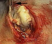
The wound is filled with pus (the correct word is purulent) that must be completely removed. Any infection that is not removed will cause the abscess to return. It is very thick and does not lend itself to easy removal.
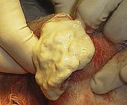
The underlying tissue that has been exposed to this infection has to be removed also. It is diseased and will be a source of further infection if it is not completely removed.
Coccidiosis comes in two different forms, intestinal coccidiosis and liver coccidiosis, depending upon the causative agent.
Both can cause rabbit diarrhea.
Acute intestinal coccidiosis can cause a life-threatening diarrhea. In some areas, the intestinal form is common, and the rabbits remain fairly healthy, while spreading the germs in their droppings. Then, when their immune systems become overwhelmed or the rabbit gets over-stressed, the symptoms flare up and the rabbit comes down with diarrhea and loss of condition.
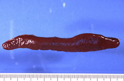
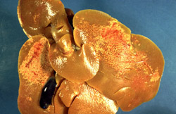
The liver form of the disease is much more concerning. Eimeria steidae, the causative protozoa, damages the bile duct and causes large white spots (pus pockets) in the liver, acute enteritis (rabbit diarrhea), loss of condition, and death. (This liver is chock full of coccidia, according to the vet report. The rabbit was unthrifty and significantly underweight for its age.)
Gastrointestinal (GI) stasis is a common cause of digestive disease in rabbits (lagomorphs). In most cases at our hospital the cause is a diet that is too low in fiber. You will sometimes read the term “ileus” when talking about this disease. Ileus occurs when the normal intestinal movement of food through the intestines, called peristalsis, stops. Normal digestion is dependent on normal peristalsis of the intestines. Peristalsis is the movement of ingesta down the stomach and intestines. When this stops GI stasis is the result.
Sometimes a hairball (also called wool block) is considered a part of this disease. In reality, over many years of treating rabbits we have learned that any hairball in the stomach is a result of GI stasis and not the cause of it. Ingesting hair is a normal part of a rabbits life, and when fed the proper food this small amount of hair passes through the digestive tract normally.
They do not get a chance to pass through the small intestines because of their location, and as a result, less nutrition is absorbed by the herbivore.
Here is a picture of the cecum taken during a routine necropsy of a rabbit. The top white arrow on the left points to just one of the 4 horizontal folds of this rabbits cecum. As you can see, it is huge and takes up a large amount of the abdominal cavity. For perspective, the arrow on the lower left points to the uterus in this female rabbit, and the arrow on the lower right points to the urinary bladder.
Another view with just a part of the cecum outside of the abdomen. Notice how the cecum has folds.
There are numerous causes to this problem:
1. Dental Disease

Rabbit teeth continuously grow. If their dental anatomy is imperfect an incisor or molar tooth can overgrow and prevent them from being able to chew their food. This will cause the GI tract to stop working and lead to stasis.
This rabbit has overgrown incisors preventing normal chewing.
2. Adhesions from prior abdominal surgery
3. Infections
4. Pain
5. Stress
6. Intestinal blockage
7. Inadequate fiber in the diet
This patient has overgrown lower incisors. They are definitely inhibiting its ability to chew. They need periodic trimming every 2-4 weeks to prevent recurrence.
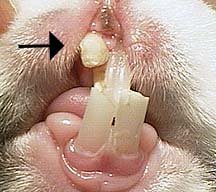
This rabbit has an upper right incisor (arrow) that has been chronically infected. We decided to remove the incisors because trimming the teeth was not solving the problem.
An acute disease characterized by a sudden outbreak of profuse, watery diarrhea, a short course and high mortality in affected animals. Predisposing factors are important and include poor sanitation, stress and sulfonamide therapy [ cocccdiosis ]. Many other species of domestic animals are also affected; therefore, interspecies transmission must be prevented. Survivors can become chronically infected and serve as carriers. In spite of the fact that rabbits in conventional rabbitries may have antibodies against the bacteria which causes this disease, outbreaks of clinical disease are usually relatively rare. However, when outbreaks do occur in commercial rabbitries, they may be devastating, with high mortality. Aside from acute deaths, chronic weight loss and intestinal stenosis occur in up to 50% of survivors .
Liver, rabbit: Small multifocal areas of necrosis.

Large intestine, rabbit: Typhylitis of the ampulla caecalis coli.

Clostridial enteropathy [ disease of the intestines caused by bacteria called clostridium ] is recognized to be a major cause of mortality in some rabbitries. This bacteria ,Clostridium spiroforme, which produces enterotoxines [ toxines in the gut lumen] is the most common clostridial pathogen associated with the enteritis complex in juvenile rabbits. Infections occur commonly in rabbitries. At necropsy of diarrheic rabbits, it was isolated from over 50% of the cases, and of those, 90% of the strains isolated were toxigenic. Clostridium perfringens also causes an enterotoxemia-like condition in young rabbits that results in bleeding enteritis in the blind sack and edema. Clostridium difficile causes infection in the large intestines in rabbits following prolonged therapy with penicillin and ampicillin. Clostridial enteropathy is recognized to be a major cause of mortality in some rabbitries. Similarly, enterotoxemia may occur due to C. spiroforme in weanling rabbits, and in adult rabbits with altered gut flora (eg. after clindamycin treatment)
Pathology:
Prominent gross lesions observed include in a large, fluid-filled edematous cecum with serosal congestion and hemorrhage and watery mucoid feces in the colon (see below). Microscopically, severe lesions include a necrotic erosive or ulcerative typhlitis with swelling and loss of enterocytes and pseudomembrane formation. The mucosa and submucosa are infiltrated with heterophils and there is submucosal edema and hemorrhage. Large Gram-positive bacilli with spores can often be observed on the mucosal surface.


The caecum is distended and the colonic content consists in a perfectly clear mucous plug.
The photo below shows a normal dropping beside a dropping encased in jelly-like mucus, indicating severe gut inflammation.

The photo below shows the difference between normal sized droppings, and those of a rabbit in the later stages of gut stasis just before he stopped passing droppings altogether. Note the small, hard and mis-shapen droppings, a clear indication that gut motility has slowed. The rabbit usually passed normal droppings but over the period of a week, the droppings got progressively smaller.

Enlarge Photo
Description:
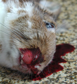
Rabbit. Severe epistaxis.
Credit: Dr. J.P. Teifke, Federal Research Institute for Animal Health, Riems, Germany
Photo ID: RHD_001
0263
Enlarge Photo
Description:
Rabbit, liver. All liver lobes are swollen, pale and have a reticular pattern.
Credit: Dr. J.P. Teifke, Federal Research Institute for Animal Health, Riems, Germany
Photo ID: RHD_002
0264
Enlarge Photo
Description:
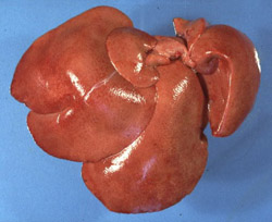
Rabbit, liver. There is a large area of pallor (necrosis) with a prominent reticular pattern.
Credit: Dr. J.P. Teifke, Federal Research Institute for Animal Health, Riems, Germany
Photo ID: RHD_003
0265
Enlarge Photo
Description:
Rabbit, lungs. The trachea is filled with foam, and the lungs are mottled and noncollapsed (severe pulmonary edema).
Credit: Dr. J.P. Teifke, Federal Research Institute for Animal Health, Riems, Germany
Photo ID: RHD_004
0266
Enlarge Photo
Description:
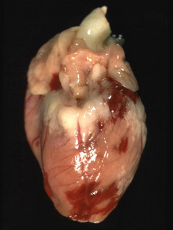
Rabbit, heart. There are multiple epicardial hemorrhages.
Credit: Dr. J.P. Teifke, Federal Research Institute for Animal Health, Riems, Germany
Photo ID: RHD_005
0267
Enlarge Photo
Description:
Rabbit, spleen. The spleen is markedly enlarged and congested.
Credit: Dr. J.P. Teifke, Federal Research Institute for Animal Health, Riems, Germany
Photo ID: RHD_006
0268
Enlarge Photo
Description:
Rabbit, kidney. There are petechiae throughout the cortex and the medulla is severely congested.
Credit: Dr. J.P. Teifke, Federal Research Institute for Animal Health, Riems, Germany
Photo ID: RHD_007
0269
Enlarge Photo
Description:
Rabbit, liver. This chronically affected liver contains pale areas of postnecrotic scarring.
Credit: Dr. J.P. Teifke, Federal Research Institute for Animal Health, Riems, Germany
Photo ID: RHD_008
0270
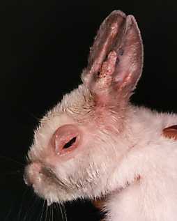
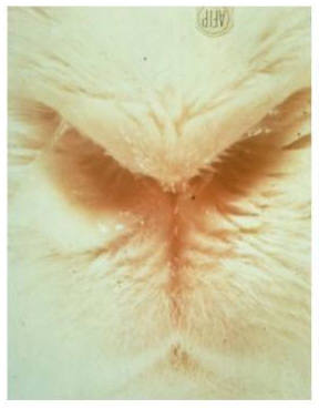
Pictured below is the vent area of a dwarf lop doe. The doe has contracted rabbit syphilis from a recent breeding.
This is also known as vent disease, vent disease in rabbits, spirochetosis, or treponematosis. The causative organism is a spirochete bacterium named Treponema paraluis cuniculi.
Mastitis is an inflammation or infection of the mammary glands, found on the rabbit's belly. Unless your doe is breeding, there is very little chance of her developing mastitis. When a rabbit is about to deliver her litter, her mammary glands will begin to fill with milk to feed the kits. The ducts in the glands open up to deliver the milk, and, of course, as soon, the kits are born, they begin nursing. Mastitis occurs as a result of bacteria moving up the duct and into the gland. Often, there is an abnormality of the gland, some trauma to th gland or nipple, or a poorly kept environment that results in the mastitis. If the mastitis is not caught right away, the kits will receive infected milk and die. You should check your doe every day while she is nursing to see if her glands appear especially swollen or red.


Ulcerated hocks are weight-bearing pressure sores on the foot of the rabbit which develop from living on hard surfaces. In severe cases the ulcers can become infected, with the infection spreading throughout the body of the rabbit and even into the bone. Ulcerated hocks are so painful that rabbits will often resort to dragging their legs behind their body to gain some relief.


The information for these diseases came from the following websites. All credit goes to them. Visit their websites for more detailed information as this is just a VERY cursory look at some of the diseases.
http://www.raising-rabbits.com/rabbit-diseases.html
http://www.lbah.com/word/rabbit-diseases/
http://www.vetnext.com/search.php?s=aan ... 77559%2080
http://www.cottontails-rescue.org.uk/gutstasis.asp,
http://www.cfsph.iastate.edu/DiseaseInf ... ic-disease,
http://www.freedomforfarmedrabbits.com/ ... sease.html,
Pasteurella Multocida
Otherwise known as Snuffles
Pasteurella multocida is a nasty little germ. When a rabbit comes down with symptoms from this bacterium, the vet will call this disease Pasteurellosis, the general term used when the rabbit’s immune system has been overwhelmed with Pasteurella multocida.
The leading researchers believe that 100% of rabbits, or at least 100% of rabbitries, have been exposed to pasteurella (Rabbit Production, Sixth Edition, pg 213). The germ is everywhere in the rabbit world; the best protection is boosting your rabbit's overall health.
This bacterium is the cause of the respiratory condition known as "snuffles." Snuffles actually describes a symptom, not exactly the illness. Snuffles are paroxysms of head-jerking sneezing that occur on a regular basis. The symptom of snuffles is almost certainly a result of pasteurella multocida. If your rabbit is sneezing, and you see ANY white sticky snot in its nose, it’s not a cold, it’s Pasteurella multocida.
Picture all those little green mucus critters on TV moving into your rabbit's lungs, suitcases and all, but with NO REMEDY.
This picture of a snotty rabbit nose is published by Tufts University.
Listen to the rabbit experts: "There’s no such thing as a ‘cold’ in rabbits," is the unequivocal statement in Rabbit Production (p. 213). "Mucopurulent nasal discharges (pus) that many people attribute to colds are almost invariably caused by P. multocida in conjunction with another bacterium called Bordatella bronchisepticum. This is not a transient condition like a cold, but one with permanent adverse effects on the animal."
Before we assume rabbit-sneezing is due to pasteurella, check to ensure the rabbit is not sneezing from environmental causes:
feed dust sniffed by the rabbit - the sneeze will be short and dry.
Drinking water in the nose - the nose might be damp on the outside tip of the nose, and the sneeze will be brief.
In the case of pasteurella: The rabbit keeps on sneezing on and off, and you notice matting on the insides of the front legs from wiping the white snot away with the front legs (they haven't figured out how to use tissues), and especially, you actually see white snot bubbles as the rabbit sneezes. Thick white snot is pasteurella multocida.
A Whole Repertoire of Trouble with Pasteurella Multocida.
Snuffles easily turns into:
Pneumonia
Metritis - infection in doe’s womb
Orchitis - infection in buck’s testicles
Colds - there are no colds, only pasteurella multocida (99% of the time)
Wry Neck - middle or inner ear infection
Weepy Eyes - eye infection closing off the tear ducts
Body abscesses - marble-sized to golf-ball sized boils, filled with thick white cheesy pus
Body ulcers - ulcerations, on skin, tongue, eyes, other locations
Bone infections - frequently associated with infected teeth
Infections in roots of teeth
I doubt this list is complete...oh yeah, death...
Symptoms
Symptoms depend on the strength (virulence) of the specific Pasteurella strain involved, which body organ(s) are involved and how long the disease is present. One of the most common symptoms is respiratory, usually manifested as a nasal discharge. When a rabbit wipes its front paws on its nose to remove the discharge the hair on the legs becomes matted. These are the symptoms that lead to the laymen’s name for this disease, snuffles.Sometimes the nasal discharge is so chronic that the fur is actually missing.
Other respiratory signs of Pasteurella include sneezing, congestion, and conjunctivitis. The tear ducts (lacrimal ducts) can become clogged with dried discharge, causing excess tearing and subsequent scalding of the skin around the eyes and face.
In addition to the respiratory tract, the bacteria can also infect the reproductive tract, the sinuses, the eyes, the ears, and the internal organs. It sometimes causes abscesses under the skin. These abscesses can become chronic and require surgery to correct. Severe cases can cause central nervous system symptoms like oscillations of the eyes (nystagmus), circling to one side, and severe tilting (wry neck or torticollis) of the head.
This rabbit has a neurologic problem from Pasteurella
Rabbits with ear infections might paw at the ears and those with internal organ infections might have poor appetites and lose weight. If the reproductive tract is infected discharge is commonly noted.
The following sections contain graphic surgical pictures, and may not be appropriate for everyone.
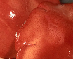
This is a healthy uterus during a routine spay (OVH). The healthy pink uterine horns are easily seen (white arrow).
The arrows point to the typical appearance of a uterus infected with Pasteurella. Cancer can also look like this.
This is what these lungs could look like on an autopsy. All the white spots correspond to the white spots on the radiograph above.
Abscesses
Rabbits have a very thick and tenacious discharge when they form an abscess, and require more care than the abscesses of most other animals. Surgical removal can be difficult, especially in the chronic cases, because the abscessed area can become extensive in nature. Multiple surgeries might be needed, and wound care at home is necessary.
This is a severe abscess on the back of a rabbit that has been anesthetized and is undergoing surgery to correct its problem. The wound has just been opened by the scalpel blade at the top left of the screen (arrow).
The wound is filled with pus (the correct word is purulent) that must be completely removed. Any infection that is not removed will cause the abscess to return. It is very thick and does not lend itself to easy removal.
The underlying tissue that has been exposed to this infection has to be removed also. It is diseased and will be a source of further infection if it is not completely removed.
Coccidiosis
Coccidiosis comes in two different forms, intestinal coccidiosis and liver coccidiosis, depending upon the causative agent.
Both can cause rabbit diarrhea.
Acute intestinal coccidiosis can cause a life-threatening diarrhea. In some areas, the intestinal form is common, and the rabbits remain fairly healthy, while spreading the germs in their droppings. Then, when their immune systems become overwhelmed or the rabbit gets over-stressed, the symptoms flare up and the rabbit comes down with diarrhea and loss of condition.
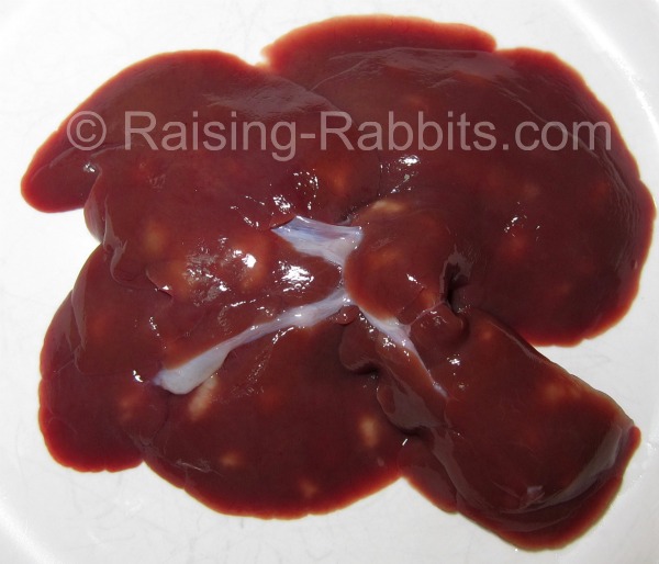
The liver form of the disease is much more concerning. Eimeria steidae, the causative protozoa, damages the bile duct and causes large white spots (pus pockets) in the liver, acute enteritis (rabbit diarrhea), loss of condition, and death. (This liver is chock full of coccidia, according to the vet report. The rabbit was unthrifty and significantly underweight for its age.)
Gastrointestinal Stasis (Hairballs)
Gastrointestinal (GI) stasis is a common cause of digestive disease in rabbits (lagomorphs). In most cases at our hospital the cause is a diet that is too low in fiber. You will sometimes read the term “ileus” when talking about this disease. Ileus occurs when the normal intestinal movement of food through the intestines, called peristalsis, stops. Normal digestion is dependent on normal peristalsis of the intestines. Peristalsis is the movement of ingesta down the stomach and intestines. When this stops GI stasis is the result.
Sometimes a hairball (also called wool block) is considered a part of this disease. In reality, over many years of treating rabbits we have learned that any hairball in the stomach is a result of GI stasis and not the cause of it. Ingesting hair is a normal part of a rabbits life, and when fed the proper food this small amount of hair passes through the digestive tract normally.
They do not get a chance to pass through the small intestines because of their location, and as a result, less nutrition is absorbed by the herbivore.
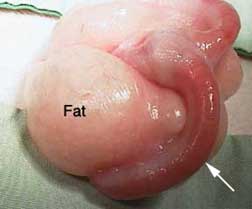
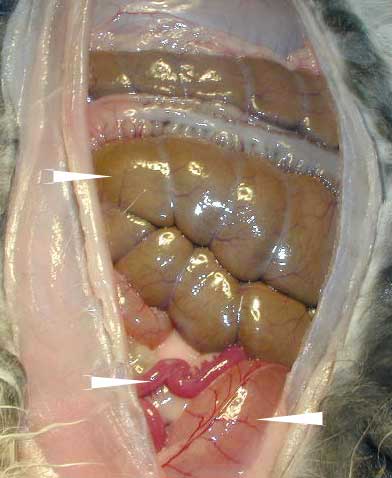
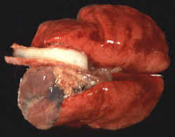
Here is a picture of the cecum taken during a routine necropsy of a rabbit. The top white arrow on the left points to just one of the 4 horizontal folds of this rabbits cecum. As you can see, it is huge and takes up a large amount of the abdominal cavity. For perspective, the arrow on the lower left points to the uterus in this female rabbit, and the arrow on the lower right points to the urinary bladder.
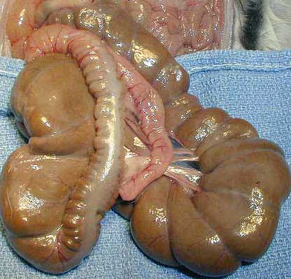
Another view with just a part of the cecum outside of the abdomen. Notice how the cecum has folds.
Cause
There are numerous causes to this problem:
1. Dental Disease
Rabbit teeth continuously grow. If their dental anatomy is imperfect an incisor or molar tooth can overgrow and prevent them from being able to chew their food. This will cause the GI tract to stop working and lead to stasis.
This rabbit has overgrown incisors preventing normal chewing.
2. Adhesions from prior abdominal surgery
3. Infections
4. Pain
5. Stress
6. Intestinal blockage
7. Inadequate fiber in the diet
Dental Disease
This patient has overgrown lower incisors. They are definitely inhibiting its ability to chew. They need periodic trimming every 2-4 weeks to prevent recurrence.
This rabbit has an upper right incisor (arrow) that has been chronically infected. We decided to remove the incisors because trimming the teeth was not solving the problem.
Tyzzer's Disease
DescriptionAn acute disease characterized by a sudden outbreak of profuse, watery diarrhea, a short course and high mortality in affected animals. Predisposing factors are important and include poor sanitation, stress and sulfonamide therapy [ cocccdiosis ]. Many other species of domestic animals are also affected; therefore, interspecies transmission must be prevented. Survivors can become chronically infected and serve as carriers. In spite of the fact that rabbits in conventional rabbitries may have antibodies against the bacteria which causes this disease, outbreaks of clinical disease are usually relatively rare. However, when outbreaks do occur in commercial rabbitries, they may be devastating, with high mortality. Aside from acute deaths, chronic weight loss and intestinal stenosis occur in up to 50% of survivors .
Liver, rabbit: Small multifocal areas of necrosis.
Large intestine, rabbit: Typhylitis of the ampulla caecalis coli.
Clostridiosis (Enterotoxemia)
Clostridial enteropathy [ disease of the intestines caused by bacteria called clostridium ] is recognized to be a major cause of mortality in some rabbitries. This bacteria ,Clostridium spiroforme, which produces enterotoxines [ toxines in the gut lumen] is the most common clostridial pathogen associated with the enteritis complex in juvenile rabbits. Infections occur commonly in rabbitries. At necropsy of diarrheic rabbits, it was isolated from over 50% of the cases, and of those, 90% of the strains isolated were toxigenic. Clostridium perfringens also causes an enterotoxemia-like condition in young rabbits that results in bleeding enteritis in the blind sack and edema. Clostridium difficile causes infection in the large intestines in rabbits following prolonged therapy with penicillin and ampicillin. Clostridial enteropathy is recognized to be a major cause of mortality in some rabbitries. Similarly, enterotoxemia may occur due to C. spiroforme in weanling rabbits, and in adult rabbits with altered gut flora (eg. after clindamycin treatment)
Pathology:
Prominent gross lesions observed include in a large, fluid-filled edematous cecum with serosal congestion and hemorrhage and watery mucoid feces in the colon (see below). Microscopically, severe lesions include a necrotic erosive or ulcerative typhlitis with swelling and loss of enterocytes and pseudomembrane formation. The mucosa and submucosa are infiltrated with heterophils and there is submucosal edema and hemorrhage. Large Gram-positive bacilli with spores can often be observed on the mucosal surface.
Mucoid enteritis
The caecum is distended and the colonic content consists in a perfectly clear mucous plug.
The photo below shows a normal dropping beside a dropping encased in jelly-like mucus, indicating severe gut inflammation.
The photo below shows the difference between normal sized droppings, and those of a rabbit in the later stages of gut stasis just before he stopped passing droppings altogether. Note the small, hard and mis-shapen droppings, a clear indication that gut motility has slowed. The rabbit usually passed normal droppings but over the period of a week, the droppings got progressively smaller.
Rabbit Hemorrhagic Disease
Enlarge Photo
Description:
Rabbit. Severe epistaxis.
Credit: Dr. J.P. Teifke, Federal Research Institute for Animal Health, Riems, Germany
Photo ID: RHD_001
0263
Enlarge Photo
Description:
Rabbit, liver. All liver lobes are swollen, pale and have a reticular pattern.
Credit: Dr. J.P. Teifke, Federal Research Institute for Animal Health, Riems, Germany
Photo ID: RHD_002
0264
Enlarge Photo
Description:
Rabbit, liver. There is a large area of pallor (necrosis) with a prominent reticular pattern.
Credit: Dr. J.P. Teifke, Federal Research Institute for Animal Health, Riems, Germany
Photo ID: RHD_003
0265
Enlarge Photo
Description:
Rabbit, lungs. The trachea is filled with foam, and the lungs are mottled and noncollapsed (severe pulmonary edema).
Credit: Dr. J.P. Teifke, Federal Research Institute for Animal Health, Riems, Germany
Photo ID: RHD_004
0266
Enlarge Photo
Description:
Rabbit, heart. There are multiple epicardial hemorrhages.
Credit: Dr. J.P. Teifke, Federal Research Institute for Animal Health, Riems, Germany
Photo ID: RHD_005
0267
Enlarge Photo
Description:
Rabbit, spleen. The spleen is markedly enlarged and congested.
Credit: Dr. J.P. Teifke, Federal Research Institute for Animal Health, Riems, Germany
Photo ID: RHD_006
0268
Enlarge Photo
Description:
Rabbit, kidney. There are petechiae throughout the cortex and the medulla is severely congested.
Credit: Dr. J.P. Teifke, Federal Research Institute for Animal Health, Riems, Germany
Photo ID: RHD_007
0269
Enlarge Photo
Description:
Rabbit, liver. This chronically affected liver contains pale areas of postnecrotic scarring.
Credit: Dr. J.P. Teifke, Federal Research Institute for Animal Health, Riems, Germany
Photo ID: RHD_008
0270
Myxomatosis
Syphilis
(Vent Disease)Pictured below is the vent area of a dwarf lop doe. The doe has contracted rabbit syphilis from a recent breeding.
This is also known as vent disease, vent disease in rabbits, spirochetosis, or treponematosis. The causative organism is a spirochete bacterium named Treponema paraluis cuniculi.
Mastitis
DescriptionMastitis is an inflammation or infection of the mammary glands, found on the rabbit's belly. Unless your doe is breeding, there is very little chance of her developing mastitis. When a rabbit is about to deliver her litter, her mammary glands will begin to fill with milk to feed the kits. The ducts in the glands open up to deliver the milk, and, of course, as soon, the kits are born, they begin nursing. Mastitis occurs as a result of bacteria moving up the duct and into the gland. Often, there is an abnormality of the gland, some trauma to th gland or nipple, or a poorly kept environment that results in the mastitis. If the mastitis is not caught right away, the kits will receive infected milk and die. You should check your doe every day while she is nursing to see if her glands appear especially swollen or red.
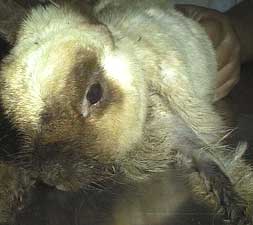
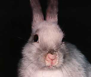
Mange
Mange is caused by contagious, parasitic mites, which burrow into the skin and ears of the rabbits and lay their eggs. This causes a great deal of itchiness and discomfort, with mites spreading rapidly from rabbit to rabbit in the severely over-crowded conditions.
Ulcerated hocks
Ulcerated hocks are weight-bearing pressure sores on the foot of the rabbit which develop from living on hard surfaces. In severe cases the ulcers can become infected, with the infection spreading throughout the body of the rabbit and even into the bone. Ulcerated hocks are so painful that rabbits will often resort to dragging their legs behind their body to gain some relief.
Ear Mites
")